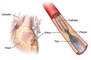
Catheter angiography utilizes a catheter, x-ray screening steer-er and an infusion of variant stuff to review blood vessels in main regions of the body for irregularities like aneurysms and illness for example plaque or atherosclerosis. The utilization of a catheter allows it likely to bring together detection and operation in a unitary process. Catheter angiography generates very comprehensive, visible and precise images of the blood vessels and may eradicate the requirement for operation.
You need to consult with doctor if there’s a likelihood if the patient is pregnant and talk about any present abnormalities, medical statements, medical advises for allergies, particularly to iodinated contrasting materials. If one is to be tranquilized, one may be instructed not to intake or drink anything for 4 to 8 hours before one’s surgical procedure. If it is there, the concerned person should make way to have someone bring him/her home. Also the concerned person should consult doctor if one has to be admitted to the hospital on short notice. Also do not wear jewelry and dress with loose, comfortable clothing.
Define Catheter Angiography?
Angiography is a least invasive medical test that assists physicians identify and handle medical situations. Angiography utilized one of three imaging technique and, in most examples, a variant material inoculation is required to make images of blood vessels in the human body.
Angiography is executed utilizing:
- CT or computed tomography
- Catheters with X-rays
- MRI or magnetic resonance imaging
In catheter angiography, a narrow plastic tube, known as a catheter, is injected into an artery through a tiny surgical cut in the skin. As soon as the catheter is controlled to the region being inspected, a variant material is inoculated through the tube and pictures are recorded utilizing a little dose of X-rays or ionizing radiation.
What are few general applications of the surgical process?
Catheter angiography is utilized to inspect blood vessels in main regions of the body, these are listed as under:
- pelvis
- brain
- heart
- arms and hands
- neck
- chest
- abdomen (for example the kidneys and liver)
- legs and feet
Doctors utilize the procedure to:
- diagnose irregularities, for example aneurysms, all in the chest and abdomen, in the aorta, or in any arteries.
- identify plaque or atherosclerotic illness in the major blood artery of the neck, which may restrict blood movement to the brain and can consequence in a stroke.
- detect a tiny aneurysm or effected arteries or veins its malformation (irregular channels between blood vessels) confined in the brain or any secondary segments of the body.
- identify atherosclerotic illness that has contracted the arteries to the legs and assists to make ready for endovascular surgery.
- diagnose illness in the arteries to the kidneys or screen blood movement to aid getting it ready for a kidney replant.
- direct interventional radiologists and doctors creating mends to infected blood vessels, for example transplanting stents or assessing a stent.
- identify damage to single or multiple arteries in the abdomen, neck, pelvis, chest, abdomen in patients after trauma
- assess arteries supply a tumor earlier to surgical process for example chemoembolization or particular inner radiation therapy.
- diagnose cut or breaking in the aorta in the abdomen or chest or its central branches.
- exhibit the scope and negative effects of the consequences coronary artery illness and schedule for a surgical process, for example a stenting and coronary bypass.
- evaluate arteries in the lungs to diagnose embolism in lungs like blood coagulate, for example those traveling from leg veins or arteriovenous defects in lungs.
- screen congenital irregularities in blood vessels, particularly arteries in kids for example defects in the heart or any blood vessels because of innate heart disease.
- study barriers of vessels.
How should We prepare?
One should intimate physician of any medications being consumed and if there are any reactions, particularly to intravenous radiocontrast variant materials. Also intimate doctor about current diseases or other medico situations.
One may be inquired to take away few or all clothes and to dress with a gown throughout the assessment. Women should always intimate physician and x-ray technologist if there is any likelihood of them being pregnant. Numerous imaging examinations are not executed throughout pregnancy in order to avoid exposure to the fetus from radiation. If an x-ray is vital, safety measures will be adopted to eradicate radiation exposure to the baby.
If a woman is breastfeeding at the time of the test, one should consult doctor how to go forward in examination. It may assists to move breast milk ahead of time and safe it on hand for utilization after contrast material has taken off from one’s body, about twenty four hours after the examination.
How does the instrument appear?
The apparatus commonly utilized for this evaluation made up of a radiographic table, single or multiple x-ray tubes and a display LCD that is situated in the evaluation room. Fluoroscopy, which changed x-rays into video pictures, is utilized to see and control advancement of the process. The video is created by the x-ray device and a identifier that is hanged over a table on which the patient resides.
The catheter utilized in angiography is an extended plastic tube about as profuse as a shore of spaghetti.
How does the process utilized?
Catheter angiography operates similar to that of a regular x-ray evaluation.
X-rays are a type of radiation similar to radio waves. X-rays can penetrate through majority of opaque bodies, as well as the human body. As soon as it is purposely targeted at the section of the body being evaluated, an x-ray machine makes a tiny break of radiation that goes through the body, capturing a picture on photographic film or an advanced detector.
Variant sections of the body soak up the x-rays in different degrees. Dense bone takes majority of the radiation whereas soft tissue, for example organs, fat and muscle permit majority of the x-rays to allow through them. As a consequence, bones are visible white on the film, soft tissue exhibits variations of gray and air takes as black.
When a variant material is brought into the bloodstream throughout the process, it sharply explicates the blood vessels being tested by allowing them exhibit bright white.
How is the process executed?
This evaluation is normally executed on an un-admitted patient. A medical practitioner will inject an intravenous (IV) line into a tiny vein in one’s arm or hand.
A tiny quantity of blood will be taken off before initiating the process to ensure that kidneys are operating and that blood will clot as per expected protocols. A tiny portion of sedative may be provided via the IV line to reduce anxiety throughout the process.
The region of the arm where the catheter will be injected is hair removed, disinfected, and deprive with regional anesthetic. The radiologist will produce a tiny incision normally some millimeters in the skin where the catheter can be injected into an artery. The catheter is then controlled through the arteries to the region to be evaluated. After the variant material is inserted through the catheter and arrives into the blood vessels being examined, numerous groups of x-rays are withdrawn. Then the catheter is taken off and the incision region is shut off by exerting stress on the region for around ten to twenty minutes or by utilizing a particular closure instrument.
When the evaluation is done, one may be inquired to hold on unless the radiologist identifies that all the mandatory pictures have been recorded. One’s intravenous line will be taken off.
A catheter angiogram may be executed within sixty minutes; on the other hand in exceptions, it may be extended for numerous hours.
What will One Encounter throughout and after the process?
Before initialization of the process, one will be inquired to make the bladder vacant. One will experience a negligible pin pierce when the needle is injected into vein for the intravenous line to be taken in. Inserting a regional anesthetic at the region where the catheter is injected may sting for a short time, but it will leave the remaining process without pain.
One will not experience the catheter in his/her artery, but when the material is inserted, one may have a sensation of coziness or a small burning experience. The utmost complex segment of the process may be lying straight for several hours. Throughout this time, one should intimate the nurse if he/she notices any pain, swelling or bleeding at the region where the catheter inserted the skin.
One may persist normal diet just after the exam. The patient will be allowed to resume all usual activities after eight to twelve hours.
Who analyze the results?
A radiologist or an expert physician particularly certified to monitor and analyze radiology tests, will interpret the pictures and forward a report to major care or concerning physician with his endorsement, who will explain the results to patient or patient’s guardian.
What are the Advantages vs. Disadvantages?
Advantages
- Angiography may eradicate the requirement for surgery. If surgery sustains mandatory, it can be executed more precisely.
- Catheter angiography exhibits an exceptional comprehensive, undoubtful and precise image of the blood vessels. This is particularly encouraging when a surgical process is being examined.
- By choosing the arteries via which the catheter transcends, it is likely to examine vessels in numerous particular body regions. Indeed, a tiny catheter may be forwarded via the extended one into a minor artery providing a tiny region of tissue or a tumor; this is known as superselective angiography.
- Apart from CT – computed tomography or MR – magnetic resonance angiography, utilize of a catheter enables it likely to involve detection and treatment in a unitary go. For instance identifying region of effected arterial contraction, subsequently chased by angioplasty and embed of a stent.
- The amount of comprehensive information by catheter angiography may not be produced with any comparitive noninvasive processes.
- No radiation stays in a human’s body subsequent to an x-ray assessment.
- X-rays normally have no demerits in the typical identification spectrum for this test.
Disadvantages
- There is a minor risk of cancer from increased and extended exposure to radiation. But, the advantage of a precise identification strikes off the danger.
- If one has an account of reaction to x-ray variant material, radiologist may counsel that he/she may consume particular drugs for twenty four hours prior to catheter angiography to reduce the danger of allergic reaction. Another alternative is to experience an alternative test that does not involve contrast material.
- If a huge quantity of x-ray contrast material emanates out within the skin where the IV is situated, skin injury can happen. If one experiences any pain in region throughout contrast material inoculation, one should quickly intimate the technologist.
- The danger of critical reaction to contrast materials that possess iodine is incredibly often, and radiology institutes are well-configured to cope up with them.
- There is a little probability that blood will make a clot adjoining the end of the catheter, hindering the artery and producing it mandatory to process to re-operate the vessel.
- If one is suffering from diabetes or kidney illness, the kidneys may be damaged because of the contrast material. In majority situations, the kidneys will resume usual operation within 5 to 7 days.
- Often, the catheter deflates the artery, resulting internal bleeding. It is probable that the catheter tip will distinguish material from the interior lining of the artery, resulting downstream obstruction in the blood vessel.
What are the restrictions of Catheter Angiography?
Patients with damaged kidney, particularly those who have diabetes, are not fit subjects for this process.
Patients who have prior reactions to variant X-ray materials are at danger of having an allergy to these materials that have iodine. If angiography is necessary, a numerous processes is utilized to minimize probability of reaction:
- One may be provided multiple dosages of a steroid drugs before time.
- Contrast material devoid of iodine may be utilized apart of quality x-ray contrast.
Catheter angiography should be executed very safely—if at all—in subjects who have a potential to lose blood.


